We are going to present several cases for you to understand patients diagnosed as atopic dermatitis previously due to:
- Abnormal IgE
- Prurigo lesions, lichenification
- Perifollicular accentuation, keratosis pilaris
- Age-specific patterns
- Early age of onset
- Personal and/ or Familial history
- Perioral lesions
Outline of Treatment if the QTT is positive
1. Treatment of the patients above 12 years old
[3] for whose body weight was more than 40KG, valyacyclovir (500 mg) 2 times a day.
Anti-allergic agents and topical corticosteroids (TCS) were prescribed to treat the dermatitis. There was no limitation for the anti-allergic agents. TCS included Medium-potency and low-potency TCS.
- Anti-allergic agents and topical corticosteroids (TCS) were prescribed to treat the dermatitis. The duration of two antiviral agents, valacyclovir (500 mg) or acyclovir (200 mg), were prescribed based on the result of the QTT.
- Antiviral agent usually prescribed for 5 to 7 days.
[1] for infants and children less than 6 years old: acyclovir granules (10 mg-15 mg/kg) 3 times a day.
[3] for whose body weight was more than 40KG, valyacyclovir (500 mg) 2 times a day.
Anti-allergic agents and topical corticosteroids (TCS) were prescribed to treat the dermatitis. There was no limitation for the anti-allergic agents. TCS included Medium-potency and low-potency TCS.
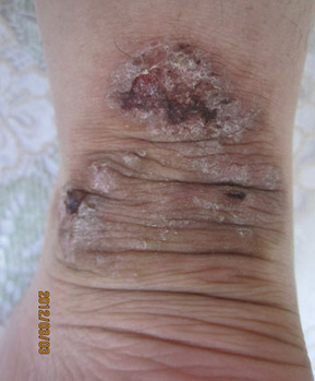
Case Number 1: Patient with plagues and prurigoes was cured by antiviral agent added into previous treatment for atopic dermatitis.
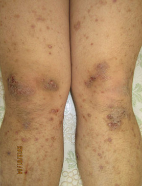
Case1-1. Prurigo type (IgE: 1272 IU/ml, HSIgG > 128). This 23-year-old man presented with itchy verrucous plaques from his face to lower extremity and with many small papules disseminated between the plaques for 2 years. Despite his history of an annual recurrence of herpes labialis for the previous 8 years, he had been treated for atopic dermatitis with anti-allergic agents and TCS, but no antiviral agent. Because the QTT from the small papules over his left knee was positive on the first visit, he was treated with 2 tablets of valacyclovir per day for 5 days combined with narrow band ultraviolet B phototherapy.
Case1-2 A large plaque with verrucous nodules above his right ankle.
Case1-3 Twelve weeks later, the verrucous nodules became flat and the plaque was smaller. There was no recurrence for 18 months after cure.
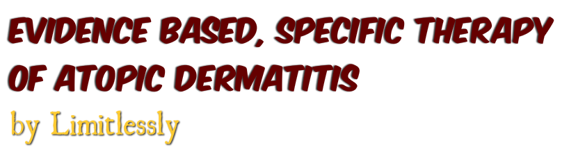
Case Number 2: Significant recovery of skin and decrease of IgE from 17220 IU/ml to 4485 IU/ml in 33 months.
This 27-year-old man presented with large itchy plaques together with lichenification 2 years ago. He had been treated for atopic dermatitis for 10 years with anti-allergic agents and TCS. The titer of his HSIgG was 78.4, and his serum IgE level was 17220 IU/ml at the beginning. The HSIgG rose to 87.2 and the IgE fell to 10660 IU/ml one year later. Clinical pictures taken 2 years after beginning antiviral therapy showed recurrent HSV labialis which was noticed for the first time in his life. There were several small red edematous nodules and plaques of various sizes on his thigh compatible with HSV-associated erythema multiforme, but there were no more large lichenified plaques.
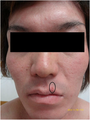
Case2-1 Erythema multiforme type (IgE: 10660 IU/ml, HS-IgG: 87.2). Clinical pictures taken 2 years after beginning antiviral therapy showed HSV labialis (cicle) which was noticed for the first time in his life.
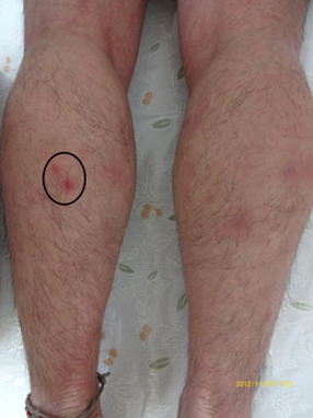
Case2-2 Two pustules (circle) surrounded by erythema on posterior aspect of his left lower leg.
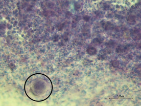
Case2-3. Intranuclear inclusion bodies were clearly observed in the nuclei of the HSV-infected cells (circle). (Magnification: ´400.)
Case Number 3: A patient with various clinical manifestations in different age.
This 21-year-old woman presented with painful erosions and vesicles on her nape and along her left hairline for 3 days. She had been treated about 3 times a year with medium potency TCS after being diagnosed with atopic dermatitis because of itchy dry skin from 2 years old to 16 years old.
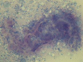
Case3-1. The QTT from a vesicle revealed HSV-infected cells as pleomorphic BCs and a giant cell with many aggregated nuclei.
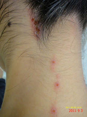
Case3-2 Three days later the roofs of the vesicles detached.
Case3-3 The lesion healed completely after Valacyclovir for 7 days
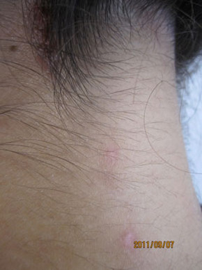
Case 3-4 This patient did not suffer any skin problem for more than 2 years. She visited again due to itchy redness, scaly desqumation over her face.
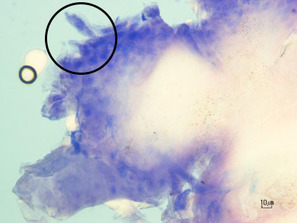
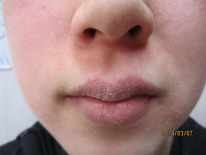
Case 3-5 Balloon degeneration with many large and irregular nuclei in the epithelium. A giant cells (circle) was also observed. This QTT was taken from perioral scaling. Her mother, grandmother, younger brother, and mother’s sister also had HSV-AD. Her mother, younger brother, and mother’s sister suffered from recurrence on the same time.
